Learn All About Dental Prophylaxis
MVZ. MSc. Andres Renato Ordoñez
Dental prophylaxis is based on the maintenance of oral hygiene at home when possible and periodontal therapy carried out by the veterinarian. The purpose of the procedure is based on the removal of all the dental plaque, as well as the polishing of each of the teeth and the revision of each of them to confirm their good condition.
Most vet practices now have some form of mechanical scaler for performing a dental prophylaxis and some practices are realising the value of having dental bases with slow and high-speed handpieces for the sectioning of teeth and the removal of bone, as well as performing more complex procedures such as endodontic therapy. Still more practices are seeing benefits in dental X-ray machine, to assist in the diagnosis of oral disease.
Oral radiology is a vital diagnostic tool in veterinary dentistry. It allows the visualization of tooth structure below the gingiva, without the superposition of the opposite jaw and other anatomical structures which may interfere with reaching a diagnosis. It offers an assessment of the periodontal and pulpal health of the tooth as well as any bony/soft tissue pathology that may be present.
The most common disease in veterinary medicine today, namely periodontal disease and also the techniques involved in tooth extractions in small animals. The disease is so common that clinicians should always examine the oral cavity thoroughly as a part of any routine health assessment.
Periodontal disease is made up of a number of plaques induced diseases involving the supporting structures of teeth. The two main forms of PD are gingivitis, the reversible inflammation of the gingiva, and periodontitis, the inflammation and irreversible destruction of the tooth’s supporting structures, namely the gingiva, the periodontal ligament, cementum and alveolar bone. Plaque accumulation leads to gingival inflammation (gingivitis). Gingivitis always precedes periodontitis, but gingivitis does not always progress to periodontitis. Periodontal Disease is caused by plaque bacteria. However, it is the interaction between plaque and the host’s immune system that can lead to the loss of alveolar bone that supports the teeth. The veterinarian’s goal for the successful management of Periodontal Disease is to minimize plaque accumulation through daily homecare and, if required, regular recalls/professional cleans.
One of the most common signs of Periodontal Disease is halitosis (oral malodour). Oral bacteria produce volatile sulphur compounds (VSC) which lead to oral malodour. VSC are also toxic to mucosal epithelium and contribute to periodontal breakdown. Other symptoms can include excessive salivation, blood-tinged saliva, dysphagia, ulceration, pain on chewing and lethargy. However, dogs with Periodontal Disease often show little or no symptoms until the disease is well advanced. This may contribute to the late presentation of these dogs to the veterinary practice, making management of their disease more difficult.
The oral examination will include inspection and palpation of the extra-oral structures including face, lips, muscles of mastication, temporomandibular joints, salivary glands, lymph nodes, maxillae and mandibles, looking for swelling, atrophy, or asymmetry. Inspection of the intra-oral structures follows, including hard and soft tissues and focusing on the dentition, gingiva, mucosa, tongue, tonsils and occlusion. On visual inspection, a dog with periodontitis may show evidence of gingival swelling, redness and an altered gingival contour around the teeth. There may also be areas with gingival recession, furcation exposures (in multirooted teeth) or purulent discharge from periodontal pockets. There can be variable amounts of plaque and calculus present, although, as a general rule, the more plaque and calculus covering the tooth surface, the more severe the disease.
Oral pathology, such as fractured teeth, may lead to altered chewing patterns with a subsequent increase in plaque, calculus and periodontitis on the affected side. The contra-lateral side can often have clean teeth with healthy gingivae. The presence and extent of plaque and calculus accumulation should be noted. The use of a plaque disclosing dye on the teeth will demonstrate to the owner the extent of the problem. However, a thorough periodontal examination can only be performed with the animal under general anaesthesia. Because periodontitis can worsen with increasing age, periodontal therapy under general anaesthesia is often performed on middle aged to geriatric pets. It is therefore important to carry out an anaesthetic risk assessment prior to embarking on what can often be a lengthy procedure.
Gingivitis is usually caused by the build up of undisturbed plaque (non specific plaque) around the tooth. The aim of treating gingivitis is to restore the affected gingival tissues back to clinical health through the thorough removal of plaque and then to maintain gingival health thereby preventing progression to periodontitis. This can often be simply accomplished by instigating such measures as homecare (e.g. daily tooth brushing) with periodic professional assessment/treatment to maintain gingival health.
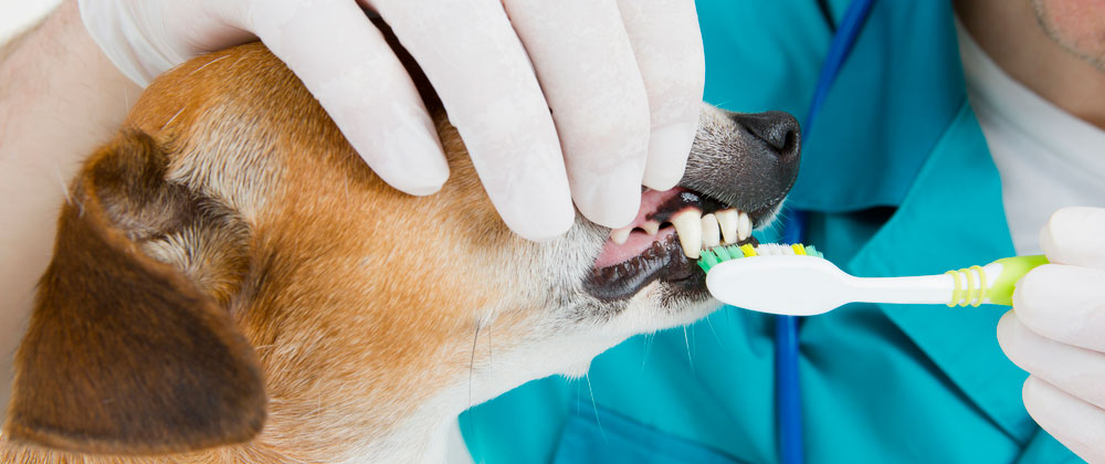
There are a large number of homecare products available to the pet owner. The gold standard for plaque removal and for the prevention of periodontal disease still remains daily tooth brushing. Without this, plaque accumulation is inevitable. Chemical agents such as chlorhexidine gluconate have also been shown to help prevent gingivitis in dogs. The use of flavoured “dog” dentifrices may aid in making the brushing experience more enjoyable for the pet.
Come, join us and we will find the best solution for your pet, we have an oral x-ray machine and of course our Fort Lauderdale animal hospital medical team is highly trained to maintain the health of our furry, Family Pet Medical Center – Family Pet Center we are always ready to give you the attention you deserve. Call today at 954-567-2500 to schedule an appointment or request emergency services.


